A deep dive into the biology and implications of inflammation
The Complete Science of Inflammation: From Molecules to Clinical Reality
Inflammation represents one of biology’s most sophisticated protective mechanisms, involving coordinated molecular cascades that can heal or harm depending on context and duration. This comprehensive analysis reveals inflammation as a tightly regulated process involving specific cellular players, molecular pathways, and resolution mechanisms—yet when dysregulated, it becomes a driver of chronic pain and disease. Recent research demonstrates that while inflammation is essential for survival, modern therapeutic approaches must navigate complex trade-offs between suppression and natural healing processes.
The evidence shows that chronic pain conditions involve distinct inflammatory patterns, anti-inflammatory treatments show surprising limitations, and dietary interventions can meaningfully modulate inflammatory processes through well-characterized molecular mechanisms. Understanding these relationships provides crucial insights for both clinical practice and personal health optimization.
What inflammation actually is at the molecular level
Inflammation is a coordinated biological response involving pattern recognition receptors (PRRs) that detect harmful stimuli, activation of specific intracellular signaling cascades, and orchestrated release of inflammatory mediators. At its core, inflammation represents the recognition of danger signals through multiple molecular pathways that converge on common transcriptional programs.
The process begins when cells detect damage-associated molecular patterns (DAMPs) like ATP, HMGB1, and S100 proteins, or pathogen-associated molecular patterns (PAMPs) such as bacterial endotoxin. These danger signals bind to pattern recognition receptors including Toll-like receptors (TLRs), triggering three major inflammatory pathways:
The NF-κB pathway serves as the master regulator, with RelA (p65) and other family members translocating to the nucleus upon activation by TNF-α, IL-1β, and other cytokines. This leads to upregulation of pro-inflammatory genes including IL-1, IL-6, IL-8, COX-2, and iNOS. The MAPK pathways (p38, JNK, ERK1/2) work in parallel, with p38 MAPK particularly important for regulating IL-1β, TNF-α, and IL-6 production. The JAK-STAT pathway provides direct translation of cytokine signals into transcriptional responses, with IFN-γ/STAT1 signaling promoting pro-inflammatory responses.
Key inflammatory mediators include cytokines (TNF-α, IL-1β, IL-6 as primary pro-inflammatory signals; IL-10, TGF-β as anti-inflammatory), chemokines (CCL2 recruiting monocytes, CXCL8 recruiting neutrophils), and lipid mediators (prostaglandins causing vasodilation and fever, leukotrienes promoting neutrophil recruitment). The complement cascade provides additional amplification through three convergent pathways producing C3a and C5a anaphylatoxins and the membrane attack complex C5b-9.
Critically, inflammation resolution is an active process mediated by specialized pro-resolving mediators (SPMs). Lipoxins, resolvins, protectins, and maresins—derived from omega-3 fatty acids—actively promote the clearance of inflammatory cells and debris while stimulating tissue repair. This resolution phase is as important as the initial inflammatory response itself.
The scientific evidence establishing inflammation’s reality
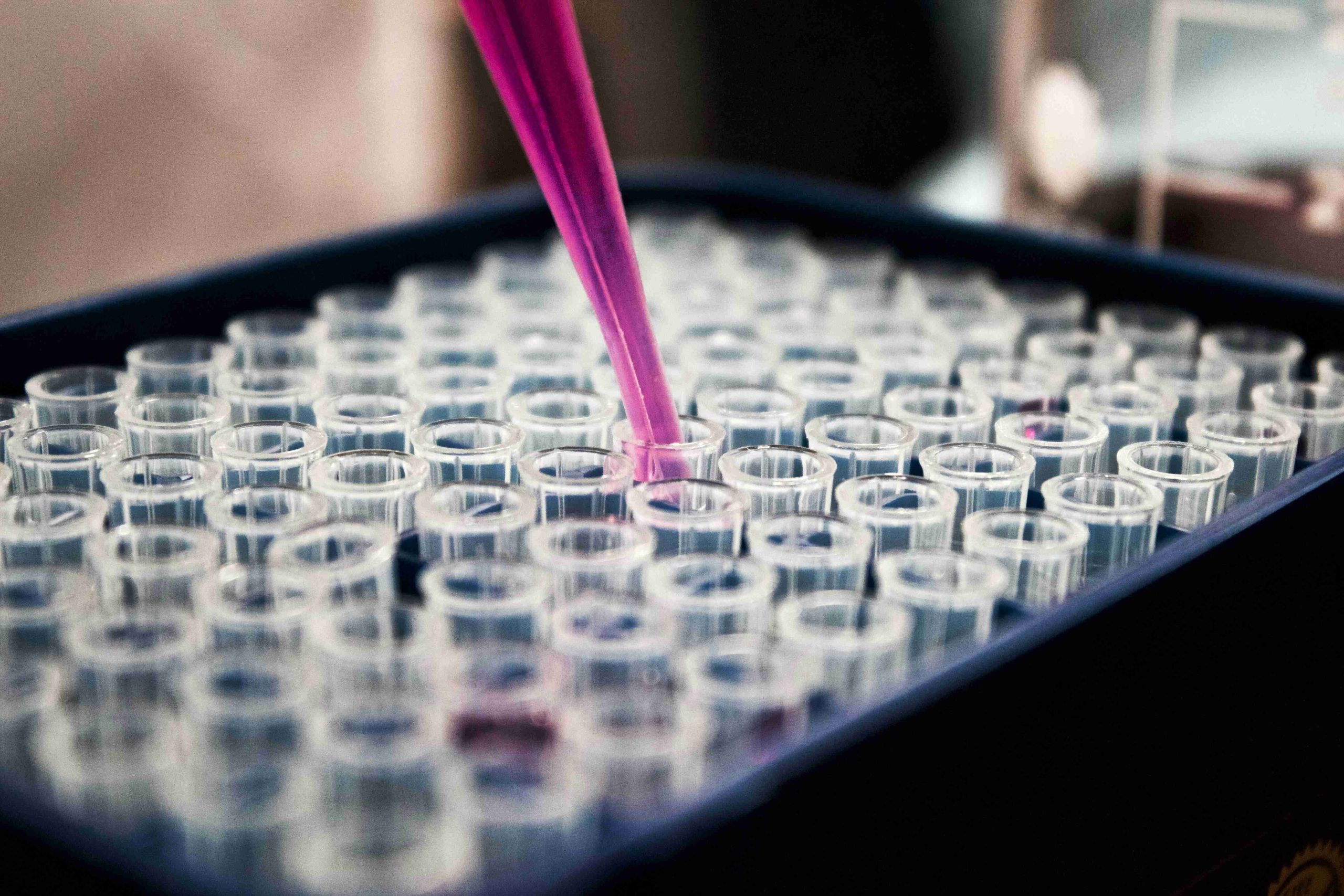
Multiple converging research methodologies have established inflammation as a measurable, reproducible biological phenomenon. In vitro studies using primary cell cultures and immortalized cell lines demonstrate consistent inflammatory responses to standardized stimuli, with cytokine measurements via ELISA, multiplex assays, and gene expression analysis via qPCR and RNA-seq providing quantitative data on inflammatory pathway activation.
Animal models including carrageenan paw edema, LPS-induced systemic inflammation, and collagen-induced arthritis show reproducible inflammatory responses that translate to human disease. Advanced imaging techniques like two-photon intravital microscopy allow real-time visualization of immune cell recruitment and behavior during inflammatory responses.
Clinical evidence includes measurable biomarkers in human studies: C-reactive protein (CRP) for systemic inflammation, pro-inflammatory cytokines (TNF-α, IL-6, IL-1β), and complement activation products. PET imaging using tracers like [11C]PBR28 now enables direct visualization of microglial activation in living human brains, providing unprecedented evidence of neuroinflammation in conditions like fibromyalgia.
Modern techniques including single-cell RNA sequencing reveal the heterogeneity of immune cell responses, while spatial transcriptomics maps inflammatory processes in tissue context. The consistency of findings across multiple methodologies, species, and experimental systems provides overwhelming evidence for inflammation as a fundamental biological process.
Inflammation’s effects on healthy individuals
In healthy people, acute inflammation follows a predictable timeline lasting hours to days, characterized by rapid NF-κB activation, transient cytokine production (TNF-α peaks at 90 minutes), and effective transition to pro-resolving mediators. This response involves coordinated vasodilation, increased vascular permeability, neutrophil recruitment, and ultimately complete resolution with debris clearance.
Chronic low-grade inflammation in otherwise healthy individuals results from lifestyle factors (poor diet, physical inactivity, psychological stress, inadequate sleep), environmental exposures (pollutants, smoking), and age-related changes termed “inflammaging.” Research demonstrates that even healthy individuals with elevated baseline inflammatory markers (CRP, IL-6, TNF-α) face increased disease risk including cardiovascular disease, diabetes, and neurodegeneration.
The gut microbiome plays a crucial role in systemic inflammation. High-fiber diets promote short-chain fatty acid-producing bacteria that have direct anti-inflammatory effects, while Western diets increase pro-inflammatory bacterial translocation and intestinal barrier dysfunction. Studies show fiber intake >25g/day associates with 66% reduction in CRP levels compared to low-fiber diets.
Age-related inflammaging involves persistent elevation of pro-inflammatory cytokines due to accumulated cellular damage, senescent cell burden, and dysregulated immune responses. This creates a pro-inflammatory environment that predisposes to multiple age-related diseases.
The inflammation-chronic pain connection revealed
Systematic reviews and meta-analyses reveal consistent elevation of pro-inflammatory cytokines across multiple chronic pain conditions, though the relationship is more complex than simple cause-and-effect.
Fibromyalgia research shows significant elevations compared to healthy controls: TNF-α (effect size 0.36), IL-6 (0.15), IL-8 (0.26), with paradoxically elevated anti-inflammatory IL-10 (0.61). Recent breakthrough research demonstrates that IgG from fibromyalgia patients can transfer pain-like symptoms to mice, suggesting an autoimmune component involving neuroinflammation through activated microglia releasing IL-1β, IL-6, TNF-α, and neurotrophic factors.
ME/CFS involves chronic immune activation similar to prolonged inflammatory responses to infection, with B cell abnormalities, gender-specific patterns (males showing altered T cell activation, females showing B cell dysfunction), and brain chemistry changes including abnormally low cerebrospinal fluid catecholamines. NIH research reveals 17-34% of patients develop ME/CFS following infections, connecting it to “long COVID” and other post-acute infection syndromes.
Migraine neuroinflammation centers on CGRP elevation during attacks, trigeminal activation releasing CGRP and substance P causing neurogenic inflammation, and meningeal inflammation with mast cell degranulation and macrophage activation. The success of CGRP antagonists in ~50% of patients provides direct evidence for inflammatory mechanisms in migraine pathophysiology.
The mechanisms linking inflammation to pain involve both peripheral and central sensitization. Inflammatory mediators directly activate nociceptors while modulating ion channels (TRPV1, TRPA1, Nav channels) and reducing activation thresholds. Centrally, spinal cord microglia and astrocytes release inflammatory cytokines that enhance excitatory transmission, reduce inhibitory signaling, and promote long-term potentiation of pain pathways through BDNF release and NMDA receptor upregulation.
Clinical reality of anti-inflammatory treatment effectiveness
The evidence reveals a striking disconnect between laboratory inflammation and clinical symptom improvement, challenging simple anti-inflammatory approaches to chronic pain.
Fibromyalgia treatment studies show consistently negative results for traditional anti-inflammatory approaches. A major JAMA meta-analysis of 224 trials (29,962 participants) found NSAIDs showed no superiority over placebo, with even approved treatments showing only small improvements (0.5-1.2 points on 0-10 pain scale) failing to reach clinically meaningful thresholds. German guidelines give negative treatment recommendations for NSAIDs in fibromyalgia due to lack of efficacy and side effect risks.
ME/CFS research reveals similar disappointments with comprehensive analysis of 56 RCTs showing no consistently effective interventions, with fewer than 5% of patients returning to pre-morbid activity levels. Low-dose naltrexone shows some promise through neuroinflammation modulation, but evidence remains limited.
Migraine represents a notable exception where mechanism-based targeting succeeds. CGRP pathway inhibitors (monoclonal antibodies like eptinezumab, erenumab) show robust efficacy in prevention with high-quality evidence, demonstrating that specific inflammatory pathway targeting can be highly effective when the right mechanism is addressed.
Chronic pain management with NSAIDs shows only modest benefits. Cochrane reviews reveal low-quality evidence for small benefit over placebo in chronic low back pain, with no superior effectiveness for sciatica. When restricted to high-quality trials, benefits are further reduced, and no evidence exists for long-term effectiveness.
The disconnect between biomarker improvements and clinical outcomes is striking. Exercise interventions in fibromyalgia show reductions in pro-inflammatory cytokines (IL-6, IL-8) despite modest clinical pain benefits. This suggests that chronic pain conditions involve altered central pain processing that may persist despite peripheral inflammation resolution, or that neuroinflammation requires different therapeutic approaches than systemic inflammation.
Why continuous anti-inflammatory drug use creates problems
Anti-inflammatory medications carry significant mechanism-based risks that accumulate with duration and dose, making continuous use problematic or dangerous.
NSAIDs disrupt essential physiological functions through COX enzyme inhibition. COX-1 inhibition reduces protective prostaglandin E2 production in the stomach, leading to increased gastric acid, decreased protective mucus, and direct mucosal damage. This increases GI bleeding risk 3-10 fold, accounting for 25% of all adverse drug reactions. Cardiovascular risks arise from disrupting the balance between pro-thrombotic thromboxane A2 and anti-thrombotic prostaglandin I2, creating a prothrombotic state with 10-50% increased risk of heart attack and stroke.
Renal toxicity occurs because kidneys depend on prostaglandins (PGE2 and PGI2) for maintaining blood flow and filtration. NSAID-induced prostaglandin suppression leads to acute kidney injury, sodium retention, and hypertension, with overall renal adverse event rates of ~18%. Recent research reveals NSAIDs also inhibit aldosterone metabolism, potentially increasing plasma aldosterone by 30-320% and contributing to cardiovascular toxicity.
Corticosteroids cause profound HPA axis suppression even with low doses for short periods. Exogenous glucocorticoids suppress hypothalamic CRH and pituitary ACTH, leading to adrenal cortex atrophy. Median prevalence of glucocorticoid-induced adrenal insufficiency is 37.4%, persisting 6-12 months after withdrawal and in some cases affecting 15% of patients three years later. Metabolic effects include diabetes (1.5-2.5 fold increased risk), osteoporosis with fractures in 30-50% of long-term users, and muscle wasting affecting both skeletal and cardiac muscle.
Biologic anti-inflammatory drugs like TNF inhibitors impair essential immune functions. Meta-analyses show statistically significant increases in serious infections and malignancies, with recent large-scale studies revealing significant increases in non-melanoma skin cancers: basal cell carcinoma (0.32% absolute risk increase) and squamous cell carcinoma (0.09% increase).
Continuous inflammation suppression disrupts natural healing processes since acute inflammation is protective and necessary for tissue repair. Recent research suggests NSAIDs may prevent the transition from acute to resolved pain by interfering with necessary inflammatory processes. Withdrawal effects include dangerous rebound phenomena: NSAID cessation increases acute myocardial infarction risk (RR 1.52) during the first month, while corticosteroid withdrawal syndrome affects patients for median 10 months with symptoms resembling adrenal insufficiency despite normal HPA function.
Glial cell inflammation drives neurological pain
Glial cells—microglia, astrocytes, and oligodendrocytes—represent a sophisticated neuroinflammatory system that fundamentally alters pain processing and contributes to chronic pain conditions through well-characterized molecular mechanisms.
Microglia serve as the brain’s resident immune cells, rapidly transforming from ramified surveillance state to amoeboid activated state upon detecting danger signals like ATP released by damaged neurons. The P2X4-BDNF-KCC2 pathway represents a crucial mechanism: microglial P2X4 receptor activation leads to BDNF release, which downregulates neuronal KCC2 chloride transporters, disrupting inhibitory signaling and promoting central sensitization.
Astrocytes maintain pain through distinct mechanisms involving JAK-STAT3 signaling triggered by IL-6, CNTF, and LIF binding to gp130 receptors. This leads to STAT3 nuclear translocation and upregulation of pro-inflammatory genes including GFAP, S100β, and complement C3. Astrocytes exist in two polarization states: A1 (neurotoxic, induced by microglial IL-1α, TNF-α, C1q) and A2 (neuroprotective, promoting repair). Reactive astrogliosis is more persistent than microglial activation and crucial for pain maintenance and spreading.
Recent single-cell RNA sequencing reveals oligodendrocytes actively participate in neuropathic pain through myelin lesion formation and production of inflammatory mediators like IL-6. They interact with other glial cells through CADM1/NRP-1/VEGFA signaling pathways, with dysfunction contributing to central sensitization.
Direct clinical evidence now exists for glial activation in human pain conditions. PET imaging using [11C]PBR28 TSPO tracer shows widespread microglial activation in fibromyalgia patients, with elevated binding in multiple brain regions including cingulate cortex correlating with fatigue levels. Human induced microglia-like cells from fibromyalgia patients demonstrate ATP hypersensitivity and enhanced TNF-α production, providing cellular validation of neuroinflammatory dysfunction.
Glial-neuron communication occurs through multiple pathways: fractalkine signaling (CX3CL1 neuronal → CX3CR1 microglial), ATP release via VNUT vesicular transporters activating surrounding microglia, and astrocytic modulation of synaptic transmission through gliotransmitter release at tripartite synapses. This creates glutamate-GABA imbalances through altered transporter function and gap junction coupling allowing inflammatory signal spreading.
Blood-brain barrier dysfunction amplifies neuroinflammation as inflammatory mediators (TNF-α, IL-1β, VEGF) from activated glia increase permeability, reduce tight junction proteins, and allow peripheral immune cell infiltration, creating positive feedback loops that sustain neuroinflammation.
Diet’s molecular influence on inflammatory processes
Rigorous research demonstrates that dietary components profoundly influence inflammatory processes through direct effects on cellular signaling pathways, with Mediterranean dietary patterns showing the strongest anti-inflammatory effects in randomized controlled trials.
Omega-3 fatty acids EPA and DHA work through multiple mechanisms: incorporation into cell membrane phospholipids alters membrane fluidity and signaling, direct NF-κB pathway inhibition prevents pro-inflammatory gene expression, and production of specialized pro-resolving mediators (resolvins, protectins, maresins) actively promotes inflammation resolution. Meta-analysis of 32 studies shows significant reductions in CRP (effect size -0.40), TNF-α (-0.23), and IL-6, but requires doses >2g/day with stronger effects at 4-6g/day.
Polyphenols demonstrate potent anti-inflammatory effects through multiple pathways. Quercetin inhibits NF-κB and reduces IL-6, TNF-α, and CRP, with 500mg/day for 8 weeks reducing rheumatoid arthritis pain and inflammation. Curcumin shows particularly robust effects with umbrella meta-analysis (21 systematic reviews, 5,870 participants) revealing CRP reduction (effect size -0.74), IL-6 reduction (-1.07), and TNF-α reduction (-1.92), with optimal dosing ≤800mg/day for <10 weeks.
The Mediterranean diet demonstrates the strongest clinical evidence for anti-inflammatory effects. The landmark PREDIMED study showed Mediterranean diet plus olive oil or nuts reduced IL-6 (mean difference -1.07 pg/mL), IL-1β (-0.46 pg/mL), and CRP (-1.00 mg/L). Meta-analysis of 22 RCTs confirms Mediterranean diet superiority over other dietary patterns for inflammatory biomarker reduction, while systematic review of 69 studies showed inverse associations with CRP in 82% of analyses.
Processed foods and added sugars consistently promote inflammatory responses through multiple mechanisms: sugar stimulates hepatic free fatty acid production generating inflammatory compounds, formation of advanced glycation end products (AGEs) from protein-sugar reactions, disruption of intestinal barrier function increasing endotoxin translocation, and activation of toll-like receptor 4 promoting cytokine release. Systematic reviews show sugar-sweetened beverage consumption positively correlates with CRP and IL-6 levels.
Gut microbiome interactions prove crucial for diet-inflammation relationships. High-fiber diets promote SCFA-producing bacteria (Bifidobacterium, Lactobacillus), with SCFAs (butyrate, propionate, acetate) having direct anti-inflammatory effects. Fiber intake >25g/day associates with 66% reduction in CRP levels, while Mediterranean diet increases beneficial bacteria while reducing pro-inflammatory Clostridium species.
Clinical significance requires substantial biomarker changes: CRP reductions >1 mg/L associate with meaningful cardiovascular risk reduction, IL-6 decreases >1 pg/mL correlate with improved insulin sensitivity, and combined dietary interventions show additive effects with Mediterranean diet reducing CRP by 1.0-2.0 mg/L and omega-3s contributing additional 0.5-1.5 mg/L reductions.
Therapeutic implications and future directions
This comprehensive analysis reveals inflammation as a double-edged biological process requiring nuanced therapeutic approaches rather than broad suppression. The evidence demonstrates that while inflammation drives many chronic conditions, simple anti-inflammatory strategies often fail because they don’t address the complex, condition-specific mechanisms involved.
Successful approaches target specific inflammatory pathways relevant to individual conditions, as demonstrated by CGRP inhibitors in migraine prevention and TNF inhibitors in inflammatory arthritis. The failure of broad anti-inflammatory approaches in fibromyalgia and ME/CFS suggests these conditions involve neuroinflammatory mechanisms distinct from peripheral inflammation or complex multi-system dysfunction beyond simple inflammatory pathways.
Dietary interventions offer the safest and most sustainable anti-inflammatory approach with Mediterranean dietary patterns, omega-3 supplementation (2-3g EPA+DHA daily), and polyphenol-rich foods providing clinically meaningful biomarker improvements without the risks associated with pharmaceutical interventions. The gut-brain-inflammation axis represents a particularly promising therapeutic target through microbiome modulation and dietary optimization.
Future therapeutic development should focus on promoting inflammation resolution rather than simply suppressing inflammatory responses, targeting specialized pro-resolving mediator pathways, and developing biomarker-guided personalized approaches that identify which patients are likely to respond to specific anti-inflammatory interventions.
The field is moving toward precision medicine approaches using PET imaging of glial activation, composite biomarker panels incorporating inflammatory and psychosocial factors, and combination therapies addressing multiple pathways simultaneously. Understanding inflammation’s complexity—from molecular mechanisms to clinical reality—provides the foundation for developing more effective, targeted therapeutic strategies that harness the body’s natural resolution pathways while minimizing the adverse effects of broad immunosuppression.
Here’s a curated bibliography of the highest-quality sources, organized by topic:
Core Inflammation Mechanisms & Pathways
Essential Reviews:
- Serhan, C.N. et al. “Specialized pro-resolving mediators: endogenous regulators of infection and inflammation.” Nature Reviews Immunology 15, 193-202 (2015)
- Chen, L. et al. “Inflammatory responses and inflammation-associated diseases in organs.” Oncotarget 9, 7204-7218 (2018)
Neuroinflammation & Glial Cell Research
Breakthrough Papers:
- “Unraveling the role of oligodendrocytes and myelin in pain.” PubMed 39162089 (2024) – Most current research on glial cells in pain
- “Single-cell sequencing reveals glial cell involvement in development of neuropathic pain via myelin sheath lesion formation in the spinal cord.” Journal of Neuroinflammation (2024)
- “Glial Purinergic Signaling in Neurodegeneration.” Frontiers in Neurology (2021)
Clinical Evidence – Chronic Pain Conditions
Fibromyalgia:
- “Brain glial activation in fibromyalgia – A multi-site positron emission tomography investigation.” Brain, Behavior, and Immunity (2018)
- “Fibromyalgia and microglial TNF-α: Translational research using human blood induced microglia-like cells.” Scientific Reports 7, 10267 (2017)
- “Inflammation, Autoimmunity, and Infection in Fibromyalgia: A Narrative Review.” PMC 11172859 (2024)
ME/CFS:
- “Molecular Mechanisms of Neuroinflammation in ME/CFS and Long COVID to Sustain Disease and Promote Relapses.” Frontiers in Neurology (2022)
- “Neuroinflammation in Patients with Chronic Fatigue Syndrome/Myalgic Encephalomyelitis: An 11C-(R)-PK11195 PET Study.” Journal of Nuclear Medicine 55, 945-950 (2014)
Treatment Evidence & Limitations
Clinical Trials:
- “Association of Therapies With Pain and Quality of Life in Patients With Fibromyalgia.” JAMA Internal Medicine (2020) – Comprehensive meta-analysis showing NSAID ineffectiveness
- “Review of pharmacological therapies in fibromyalgia syndrome.” Arthritis Research & Therapy (2014)
Cochrane Reviews:
- “Non-steroidal anti-inflammatory drugs for chronic low back pain.” Cochrane Database (Updated reviews)
- “Non-steroidal anti-inflammatory drugs for low back pain with sciatica.” Cochrane Database
Diet & Inflammation Research
Key Meta-Analyses:
- “Effects of Dietary Patterns on Biomarkers of Inflammation and Immune Responses: A Systematic Review and Meta-Analysis of Randomized Controlled Trials.” ScienceDirect (2022)
- “Mediterranean Diet as a Tool to Combat Inflammation and Chronic Diseases. An Overview.” PMC 7400632 (2020)
- “The Immunomodulatory and Anti-Inflammatory Role of Polyphenols.” PMC 6266803 (2018)
Biomarkers & Measurement
Recent Advances:
- “Biomarkers for Chronic Pain: Significance and Summary of Recent Advances.” PMC 9663208 (2022)
- “Innovations in acute and chronic pain biomarkers: enhancing diagnosis and personalized therapy.” PMC 11877092 (2024)
Drug Safety & Mechanisms
Authoritative Sources:
- “Risk of Adverse Events After Anti-TNF Treatment for Inflammatory Rheumatological Disease. A Meta-Analysis.” PMC 8591221 (2021)
- “Pathophysiological aspects of nephropathy caused by non-steroidal anti-inflammatory drugs.” PMC 6534025 (2019)
- “Glucocorticoid Therapy and Adrenal Suppression.” NCBI Bookshelf NBK279156

